Chapter 1 : The Cells of the CNS

Brain: Contents Page
The Cells of the CNS.          Topics :     

Physiology of Glial Cells

Overview of Glial Cells in the Central and Peripheral Nervous Systems

Glial Cells occur in approximately the same numbers as neurones in most parts of the CNS.

There are four main functions of glial cells:

  1. To surround neurones and hold them in place,
  2. To supply nutrients and oxygen to neurones,
  3. To insulate one neurone from another,
  4. To destroy pathogens and remove dead neurones.


Astrocytes have processes that contact nerves cells, e.g. at synapses or nodes of Ranvier, and others that are in contact with blood vessels. This structure is thought to help regulate the supply of nutrients to the neurones and maintain local homeostasis of the environment of the neurones..

Oligodendrocytes provide the myelin wrapping of large axons in the CNS, and are therefore responsible for maintaining a high speed of conduction in central nervous pathways.

Microglia are the dormant phagocytes of the central nervous system, and can be activated to become phagocytes in these tissues

Ependymal cells are ciliated cells that line the ventricular system.

*


Variety of Glial Cell Structures Astrocytes
Oligodendrocytes
Microglia
Ependymal Cells
Satellite Cells
Tanycytes

Docstoc

Top


Astrocytes and the Physiology of the Blood Brain Barier

It has been know for over a century that if certain dyes were injected into the bloodstream of an animal, the tissues of the whole body EXCEPT the brain and spinal cord would be coloured.

Similarly if the dye was injected into the brain, the brain would be coloured, but the colour would not cross into the body.

So the concept of a "Blood-Brain Barrier" (BBB) which prevents materials in the blood from entering the brain, and vice versa, came into existence. More recently, the cellular structures associated with this barrier have become more understood.

The main functions of the blood-brain barrier are to protect the brain from circulating organisms or chemicals that might interfere with brain function and to maintain a stable environment for the neurones to perform their functions.

Interference with brain function could arise from entry of neurotransmitters, metabolites, osmolality, hormones and toxins, and of organisms such as bacteria and viruses; so it is important that these are not allowed tohave access to neurones and synapses.


Capillaries are formed of a tube of endothelial cells in fairly close contact, so that, in most parts of the body, large molecules cannot cross the normal endothelium . There are exceptions, such as the glomerulus of the kidney and the capillaries of the bone marrow, where the endothelium is fenestrated- i.e has 'windows', which make it leaky.

Brain Capillaries are made of endothelial cells that are tightly bound together, at 'tight junctions', and these vessels prevent large molecules from crossing to the extracellular fluid of the brain. Outside the endothelial cells is the basement membrane which may be thickened in brain capillaries.

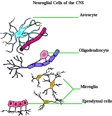
Role of Astrocytes. In addition to tight junctions and a thickend basement membrane, a specialised type of glial cell, the astrocyte, controls the environment of neurones within the brain.

One of its functions is to be selective in the transfer of materials from the blood to the neurones of the brain. It does this by having processes which terminate in end feet on the outside of the capillaries, and others on the neurone. As a result the astrocyte is the highly selective link between the neurone and the blood. In addition to their barrier function, astrocytes appear to play an important part in transporting ions and other chemicals from the brain to the blood.

Astrocytes are also involved in disposing of unwanted neurotransmitters from the extracellular fluid around neurones and synapses (see below).

Astrocytes These have numerous projections that anchor neurons to their blood supply. They appear to regulate the external environment of neurons by a selective movement of ions (e.g. potassium) and other chemicals.

They are able to take up various chemical released at synapses and help to recycle them; one example would be the neurotransmitters released during synaptic transmission.

Astrocytes are believed to be an essential part of the blood–brain barrier, and may release substances that regulate local blood flow, so that local blood flow is matched to the metabolic activity of neurones in that area.

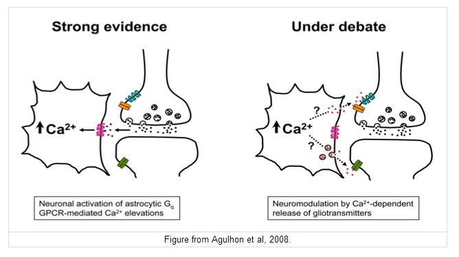
Astrocytes are connected to each other by gap junctions, and communicate using calcium and other chemical messengers.


*

Some Properties of the BBB.

Like may biological membranes, the blood brain barrier is semipermeable; it allows some molecules to cross while repelling others. Generally speaking, charged molecules are repelled, and are only transported by specific transport mechanisms; lipid-soluble molecules that can dissolve in cell membranes may be able to cross, e.g. the barbiturate group of drugs.

Small molecules such as oxygen, carbon dioxide and glucose appear to cross either by diffusion of by some specific transport mechanism.

It is also recognised that the BBB is not fully formed at birth, so brain development is particularly vulnerable to pathophysiological events in infants.

The BBB can be breached in certain pathophysiologiucal states including: hypertension, hyperosmolality, infection, trauma, ischaemia, inflammation, radiation, and raised intracranial pressure. Should the BBB be breached, microglia are activated and become phagocytic in nature, removing cellular debris, invading organisms etc.


Some Exceptions:

Some sites within the brain however do monitor chemical concentrations within the blood. One example is the level of hormones that are sensed by the hypothalamus.

In parts of the brain where monitoring of blood chemistry takes place, the blood brain barrier is defective, and it is known that this defect is present at a number of sites around the cerebro-ventricular system. These sites are given the name circumventricular organs


The circumventricular organs allow the brain to monitor aspects of blood chemistry. They occur mainly around the Third Ventricle, where several areas are involved in monitoring osmolality and hormone levels, and in the medulla, where toxic substances can enter the brain and give rise to vomiting 'the vomiting centre is close to the area postrema.

All of the circumventricular organs have an extensive blood supply and fenestrated capillaries cause them to be leaky at these sites. These sites have special functions including the regulation of body fluid balance, cardiovascular control, immune responses, thirst, feeding behaviour and sexual behaviour.


Role of Astrocytes in Cerebral Blood Flow

Cerebral Blood Flow
Local Blood Flow in the Brain
Links between Neuronal Metabolism and Local Blood Flow


Armin Kübelbeck

Top

Myelination in the Central and Peripheral Nervous Systems

Oligodendrocytes

Oligodendrocytes are cells that coat axons in the CNS with their cell membrane forming a myelin sheath, that insulates the axon and is essential for fast transmission of nerve impulses. Defects in this system occur in demyelinating diseases such as Multiple Sclerosis.

In contrast to the myelin of the peripheral nervous system, one oligodendrocyte can provide a myelin sheath for several axons.

*

Schwann Cells and Myelination

In the peripheral nervous system, Schwann cells provide the myelin sheath of axons which they surround.

Myelin forms by the Schwann cell wrapping itself around the axon many times. There are therefore many layers of cell membranes around the axon, and their high electrical resistance causes action potentials at each node of Ranvier to jump to the next node. This is called Saltatory Conduction.

Schwann cells may also have phagocytic activity and clear cellular debris that allows for regrowth of PNS neurons after nerve transection..


Schwann cells are also involved in the support of unmyelinated nerve axons. In this situation, no myelin sheath is formed, but several axons are surrounded by the Schwann cell, forming bundles of unmyelinated axons known as Remak Bundles.

*


Demyelinating Diseases Multiple Sclerosis

Wikipedia
Openi.com

Top


Glial Cells and the Metabolism of Neurotransmitters in the CNS

The amino acid Glutamate is the most important excitatory neurotransmitter in the CNS. It is synthesised from glutamine within the axon terminals and stored within vesicles in synaptic boutons.

After its release into the synapse glutamate is transported into astrocytes which convert it into glutamine, which is recycled by the nerve endings.


Glutamate Synthesis

Neurons synthesise glutamate from glutamine.

Glutamate is synthesised within glutamatergic nerve endings, but after release, it needs to be removed from the synapse quickly because accumulation of extracellular glutamate is associated with neuronal toxicity (see below).

The glutamate-glutamine cycle involves astrocytes

The glutamate-glutamine cycle is recognised as an important metabolic pathway for mopping up excess glutamate from the synapse.

It is a mechanism whereby glutamate released at synapses can transported into adjacent astrocytes (and neurones) using a family of transporters - the Excitatory Amino Acid Transporters (EAAT) - and converted glutamine within the astrocyte.

This glutamine can be recycled and passed to the nerve endings for the synthesis of glutamate. Excess glutamine is transported away from the brain in the blood stream.

*

Astrocytes and GABA Metabolism

A similar process occurs for GABA, a major inhibitory neurotransmitter within the CNS. Following its release into the synaptic cleft it is taken up by astrocytes and converted into glutamine which is reused (see the right hand side of the diagram).


Interactions between Glia and Neurones; Gliotransmitters   Top

The diagram opposite shows that neurotransmitters released by nerve can act on glial cells - it is known that neurotransmitters can initiate calcium entry to astrocytes, and that the changes in membrane potential can be transmitter amongst a group of astrocytes because of the presence of gap junctions.

In addition it is thought that substances released from glial cells can alter the sensitivity of post-synaptic neurones to neurotransmitters. The diagram (right) indicates a possible scenario for glial-neuronal interactions, and there is increasing evidence for the existence of gliotransmitters, i.e. neuromodulators that influence the adjacent synapses. There are several contenders for the role of gliotransmission including glutamate and ATP.


www.bioscience.org

Top


Brain: Contents Page

The Cells of the CNS.          Topics :     

© HumanPhysiology.Academy 2014-2015